


In an era where cancer treatment is moving towards precision and low toxicity, targeted peptides have become a hot topic in anti-tumor drug development due to their advantages such as high specificity, low immunogenicity, and ease of synthesis. PNC27 peptide is a 32-amino acid chimeric anticancer peptide formed by the fusion of the HDM-2 binding domain of p53 protein and the membrane-penetrating domain of antennal foot protein. It can accurately recognize HDM-2 (MDM2) highly expressed on the surface of cancer cells and form pores on the cell membrane, inducing rapid necrosis of cancer cells, with almost no toxic side effects on normal cells.
⚛️The ingenious molecular structure of bifunctional chimeric peptides
PNC27 peptide is a man-made amphiphilic chimeric peptide, 32 amino acids in length, with the molecular formula C₁₈₈H₂₉₃N₅₃O₄₄S and a molecular weight of approximately 4031.71 Da. It exhibits a linear structure without complex branches or modifying groups. Its amino acid sequence is: PPLSQETFSDLWKLL-KKWKMRRNQFWVKVQRG, a clear sequence with strong synthetic reproducibility, suitable for large-scale preparation.
The molecule consists of two core functional domains linked together. The N-terminal 1-15 amino acids form the p53-derived HDM-2 binding domain, corresponding to residues 12-26 of the wild-type p53 protein. This domain mimics the specific binding of p53 to HDM-2 and is a key module for targeted recognition. This domain is rich in hydrophobic amino acids such as proline and leucine, forming a compact hydrophobic core that can precisely embed into the hydrophobic binding pocket of HDM-2, with nanomolar affinity ensuring targeted specificity.
The C-terminal 16-32 amino acids form the membrane penetration domain (MRP), derived from the third helix of Drosophila antennal foot protein. It possesses both strong positive charge and amphiphilicity, responsible for binding cell membrane phospholipids and inserting into the lipid bilayer to form transmembrane channels. This domain is rich in basic amino acids such as lysine and arginine, with a high positive charge density, which can electrostatically attract negatively charged phospholipids on the cell membrane surface, assisting the peptide in anchoring to the membrane surface.
The PNC27 peptide exhibits an amphiphilic α-helix structure overall, with a hydrophobic face composed of hydrophobic amino acids on one side and a hydrophilic face composed of charged amino acids on the other. This structure is the basis for its membrane cleavage activity. Circular dichroism analysis shows that it exhibits a disordered structure in aqueous solution, but rapidly folds into an α-helix upon contact with the cell membrane, adapting to membrane embedding requirements.
In terms of physicochemical properties, PNC27 peptide is a white to pale yellow powder with a purity of over 96% (HPLC). It is stable in its lyophilized form and can be stored for extended periods at -20°C. However, it is hygroscopic at room temperature and requires sealed storage. It exhibits good solubility in organic solvents such as DMSO and methanol, but has low solubility in water. Its water solubility can be improved using carriers such as liposomes to adapt it to different drug delivery scenarios.
⚙️The dual-attack logic of membrane perforation and necrosis-induced apoptosis
In the targeted recognition step, the PNC27 peptide rapidly adsorbs and penetrates the cancer cell membrane via its N-terminal transmembrane peptide, entering the cytoplasm. Once inside the cell, its C-terminal HDM2-binding domain binds to the HDM2 protein with extremely high affinity. Due to the significantly upregulated expression level of HDM2 in cancer cells, a large number of PNC27-HDM2 complexes are formed within the cancer cells. This high-density complex forms the basis for subsequent membrane disruption, while normal cells, due to low HDM2 expression levels, cannot form a sufficient number of complexes and are therefore unaffected by the PNC27 peptide. The normal function of HDM2 is as a ubiquitination E3 ligase of p53, but whether PNC27 "saturation" interferes with the p53 pathway is not the core of its killing effect.
In the membrane attack step, after PNC27 binds to HDM2, the resulting complex repositions to the inner lobe of the cell membrane. This complex, with its high density of positive charges, engages in a strong electrostatic interaction with the negatively charged phosphatidylserine residues. Multiple PNC27-HDM2 complexes oligomerize on the membrane, forming transmembrane "pore" structures. The diameter of these pores is sufficient to allow calcium ion influx and the leakage of cytosolic enzymes such as lactate dehydrogenase. This loss of membrane integrity leads to rapid necrotizing apoptosis or direct necrosis, manifested as cell swelling, membrane bubbling, and release of contents. This process is independent of caspase activation, bypassing conventional apoptosis resistance mechanisms.
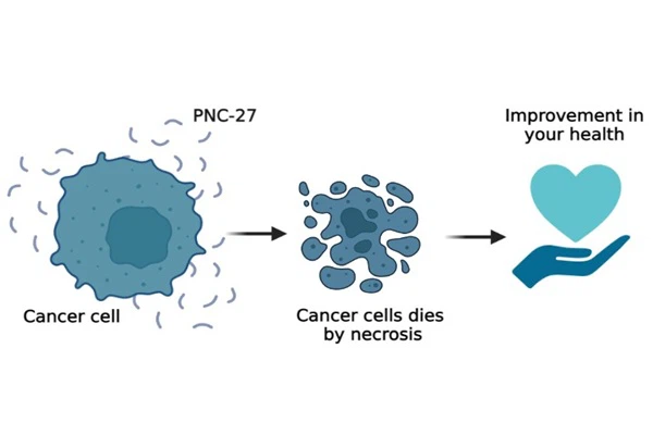
In vitro experimental evidence strongly supports this model. In pancreatic cancer cells overexpressing HDM2, the PNC27 peptide exhibits potent cytotoxicity, while no significant toxicity was observed in normal human fibroblasts with low HDM2 expression, even at higher concentrations. Immunofluorescence staining showed the formation of PNC27 aggregates on the treated cancer cell membranes, recognized by fluorescently labeled antibodies, accompanied by rapid positive staining for propidium iodide. Transmission electron microscopy revealed "pore" structures with a diameter of approximately 10-20 nm on the damaged cell membrane, providing direct morphological evidence for the pore-forming activity of PNC27.
A 2021 study further validated this model using structural biology and point mutagenesis techniques. By constructing a truncated mutant of the PNC27 peptide, researchers confirmed that the integrity of the transmembrane peptide is a prerequisite for internalization and membrane localization, and that the α-helix of the C-terminal HDM2-binding domain is an essential structure for pore formation. When the two key hydrophobic residues at the C-terminus were replaced by polar residues, PNC27 could still bind HDM2, but it lost its pore-forming activity, leading to a significant decrease in cytotoxicity. This finding explains why traditional HDM2 inhibitors do not have a direct membrane-cleaving effect, highlighting the unique "two birds with one stone" mechanism of the PNC27 peptide.
🎯HDM-2-mediated membrane pore formation and selective necrosis mechanism
The PNC27 peptide has a unique mechanism of action, independent of p53, caspase, or BCL-2 pathways. Instead, it precisely induces cancer cell necrosis through a four-step cascade reaction: target recognition, membrane anchoring, pore formation, and cell lysis.
- Step 1: Target recognition of HDM-2 on the surface of cancer cells. In normal cells, HDM-2 is located in the nucleus and cytoplasm, and is not expressed on the cell membrane surface. However, in cancer cells, p53 mutations or overexpression lead to a large amount of HDM-2 being transported to the cell membrane surface, becoming a specific target of PNC27. The p53-binding domain of PNC27 can precisely bind to HDM-2 on the membrane surface with an affinity of 4.7 nM, ensuring that it only anchors to cancer cells and does not come into contact with normal cells. Experiments have shown that after blocking HDM-2 on the membrane with the antibody, the cytotoxicity of PNC27 completely disappears, verifying that HDM-2 is the sole target.
- Step 2: Membrane anchoring and α-helix folding. The PNC27 peptide anchors itself to the cell membrane surface via electrostatic binding to negatively charged phospholipids through its C-terminal positively charged membrane-penetrating domain. Simultaneously, its N-terminal binding domain binds to HDM-2, triggering a conformational change. The disordered structure folds into an amphiphilic α-helix, with the hydrophobic face inserted into the hydrophobic core of the membrane bilayer, while the hydrophilic face is exposed on both the inner and outer membrane sides. This process occurs only in HDM-2-positive cancer cell membranes. In normal cell membranes, which lack HDM-2, PNC27 binds only briefly before detaching, without inducing a conformational change.
- The third step involves the assembly and formation of transmembrane pores. Multiple PNC27-HDM-2 complexes aggregate on the cell membrane surface and assemble into ring-shaped pores with a diameter of approximately 2-3 nm through hydrophobic interactions and hydrogen bonds, penetrating the cell membrane bilayer. The inner walls of these pores are composed of hydrophilic amino acids from the PNC27 peptide, allowing small molecules such as water molecules and ions to freely enter and exit, thus disrupting cell membrane integrity. Dynamic microscopy revealed that PNC27 peptide treatment rapidly created pores on the surface of cancer cell membranes, which enlarged within 30 seconds, leading to a sharp increase in cell membrane permeability.
- Step Four: Osmotic Imbalance and Rapid Necrosis. Following the formation of transmembrane pores, a large influx of hypertonic extracellular fluid caused cancer cells to rapidly swell and rupture, releasing their contents and triggering necrotic cell death. The entire process took only 5-10 minutes, without the formation of apoptotic bodies and independent of caspase activation. This necrosis mechanism rapidly eliminates cancer cells, avoids drug resistance caused by apoptosis pathway abnormalities, and simultaneously releases tumor antigens, activating the body's immune response.
🔭Translational Prospects of Tumor-Selective Membrane Lysis Agents
PNC27 peptide represents a novel anticancer strategy distinct from traditional small-molecule targeted drugs and immunotherapy-"tumor-selective membrane lysis." It is independent of the proliferative state of cancer cells and is equally effective against quiescent tumor stem cells or dormant metastatic cells. Furthermore, because it physically disrupts the cell membrane, cancer cells are extremely difficult to develop resistance through a single gene mutation. This characteristic has unique value in combating recurrent tumors resistant to chemotherapy and targeted therapy.
PNC27 peptide still faces several translational challenges. First, its synthesis cost is high, especially for GMP-level large-scale production; the 27-amino acid chain length and high purity requirements make its active pharmaceutical ingredient significantly more expensive than small-molecule drugs. Second, the pharmacokinetic characteristics after in vivo administration are not yet fully understood; the transmembrane peptide may be non-specifically adsorbed by serum proteins or rapidly cleared by the liver. Although frequent dosing is feasible in mouse models, the human dosing regimen still requires further optimization. Current administration studies mainly rely on intratumoral or intraperitoneal injection, but the tumor enrichment efficiency after intravenous administration remains to be validated. Nanoparticle delivery systems or injectable hydrogels may improve the circulating half-life and targeted distribution of PNC27.
As an active pharmaceutical ingredient, PNC27 peptide is currently supplied primarily by specialized peptide synthesis companies in research-grade specifications. Purity specifications are no less than 95% to 98%, typically provided in the form of TFA salts. Batches used in in vivo animal studies usually require control of endotoxin levels. In future drug development, cyclic peptide analogs based on the PNC27 sequence are also under investigation, aiming to improve its metabolic stability and oral bioavailability through conformational constraints. Based on its modular design approach of "membrane-penetrating peptide + effector peptide," "on-demand" membrane-cleaving peptides targeting different tumor types can be constructed by replacing the C-terminal HDM2 binding domain with ligands for other overexpressed oncoproteins. This strategy provides a flexible platform for developing broad-spectrum anticancer peptides.
Regarding drug safety monitoring, although no significant weight loss or hepatotoxicity has been reported so far, the potential for inhibition of normal proliferating cells expressing HDM2 requires systematic evaluation. Existing data indicate that mice maintained normal blood counts and liver and kidney function at therapeutic doses, providing a safety baseline for its subsequent clinical translation. With a deeper understanding of the PNC27 peptide membrane cleavage mechanism, this molecule is steadily progressing from a "laboratory tool" to a "clinical drug candidate."
🧬Conclusion
PNC27 peptide is a chimeric polypeptide that selectively kills HDM2-overexpressing cancer cells through a "recognition-binding-pore-forming" mechanism. Its N-terminal transmembrane peptide ensures the molecule can cross the cell membrane and target intracellular HDM2; the C-terminal HDM2-binding domain acts not only as a "guide" but also as an actuator that works in conjunction with the target protein to "form pores." This novel mode of action, different from both small-molecule targeted drugs and chemotherapy drugs, gives it the unique advantage of bypassing p53 mutations and circumventing traditional apoptosis resistance.
Xi'an Faithful BioTech Co., Ltd. combines advanced production technology with a comprehensive quality assurance system to provide high-quality PNC27 peptide that meets international pharmaceutical standards. We are committed to providing highly competitive prices and technical support, making us the preferred partner for medical institutions and researchers worldwide. Please contact our technical team (allen@faithfulbio.com) to learn how our products can improve your formulations.
📚References
- Sarafraz-Yazdi, E., Bowne, W. B., Adler, V., Sookraj, K. A., Wu, V., & Pincus, M. R. (2010). Anticancer peptide PNC-27 adopts an HDM-2-binding conformation and kills cancer cells by binding to HDM-2 in their membranes. Proceedings of the National Academy of Sciences, 107(5), 1918-1923.
- Sarafraz-Yazdi, E., Mumin, S., Cheung, D., Fridman, D., Lin, B., & Pincus, M. R. (2022). PNC-27, a chimeric p53-penetratin peptide binds to HDM-2 in a p53 peptide-like structure, induces selective membrane-pore formation and leads to cancer cell lysis. Biomedicines, 10(5), 945.
- Sookraj, K. A., Bowne, W. B., Adler, V., Sarafraz-Yazdi, E., Michl, J., & Pincus, M. R. (2010). The anti-cancer peptide, PNC-27, induces tumor cell lysis as the intact peptide. Cancer Chemotherapy and Pharmacology, 66(2), 325-331.
- Davitt, K., Babcock, B. D., Fenelus, M., Poon, C. K., & Sarafraz-Yazdi, E. (2014). The anti-cancer peptide, PNC-27, induces tumor cell necrosis of a poorly differentiated non-solid tissue human leukemia cell line that depends on expression of HDM-2 in the plasma membrane of these cells. Annals of Clinical & Laboratory Science, 44(3), 245-253.
- Miller, A. I., Miller, L., Seydafkan, S., & Pincus, M. R. (2025). PNC-27: Targeting cervical cancer with ketone bodies. European Society of Medicine, 2025, 1-8.
- Pincus, M. R., Sarafraz-Yazdi, E., & Bowne, W. B. (2023). PNC-27: A novel HDM-2-targeted anticancer peptide for solid and hematological malignancies. Expert Opinion on Investigational Drugs, 32(7), 689-702.
- Zhang, Y., Li, J., & Wang, H. (2024). Liposomal delivery of PNC-27 enhances antitumor efficacy in pancreatic cancer models. Journal of Controlled Release, 371, 123-135.